Microscopes in Action
Symbiotic Salamanders
Exploring symbiosis at the cellular level

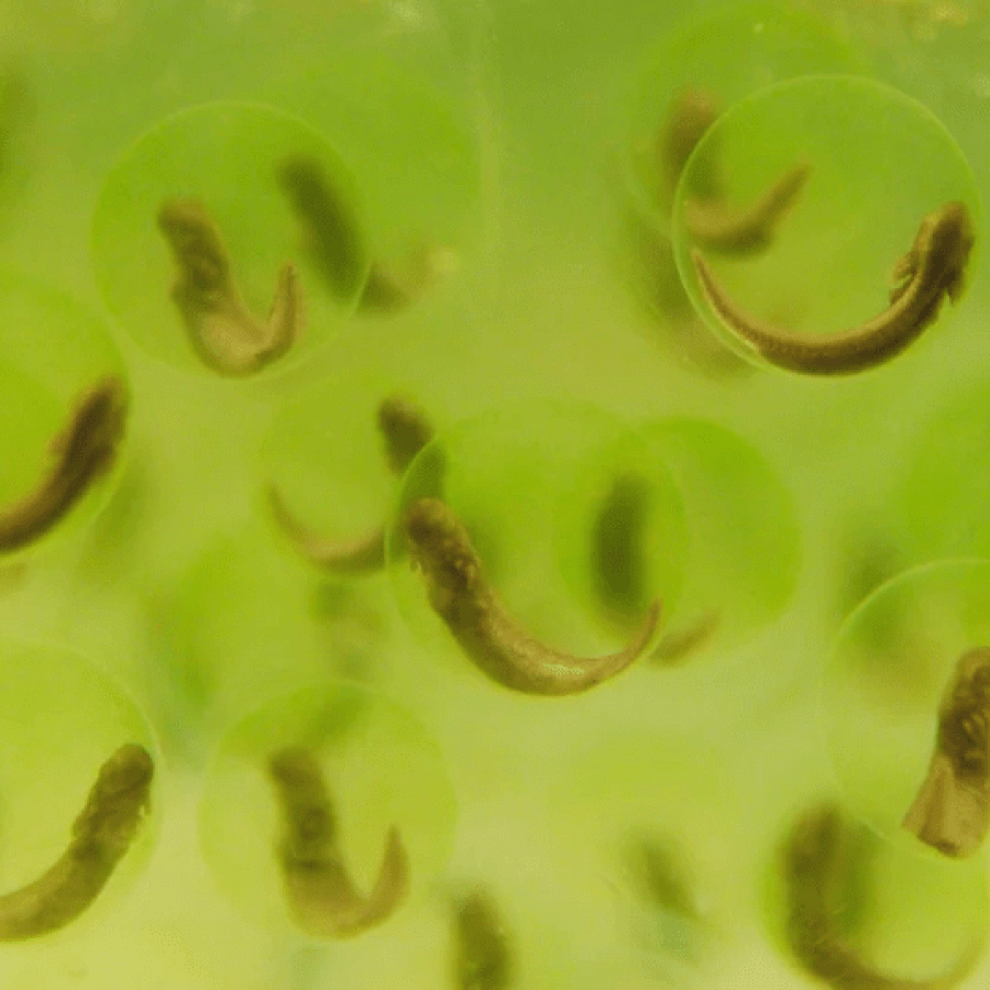
The eggs of the yellow spotted salamander, a species native to Nova Scotia, contain many green plant cells called algae. As shown in the photograph to the right, the salamander embryo (indicated by the back arrow) is present within the egg capsule and is surrounded by green algal cells that are too small to be seen individually.
The small algae promote growth of the salamander embryo while the embryo provides nutrients and protection for the algae. This relationship is called symbiotic as it helps both partners to thrive.
Researchers in the Bishop lab @StFX have been studying this relationship at the cellular level using a variety of different microscopes to view the small algal cells. The image below, taken using the Scanning Electron Microscope, clearly shows the algae clustered on inside wall of the egg capsule.

The light microscope was used to view a section of the interior structure of the algal cells, as shown below:

The inner structure of the algal cell and its attachment to the egg capsule wall is revealed in the transmission electron micrograph below:

Visit the Bishop Lab to learn more about this research
Seaweed Shedding
Getting rid of pests
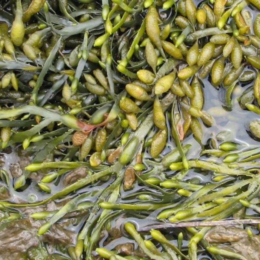
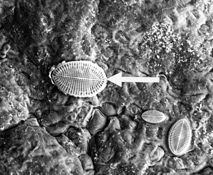
Seaweed is present around the coast of Nova Scotia. Several different organisms, such as the diatom indicated here by the white arrow, settle upon the upper leaves of the seaweed and block out the sun’s rays.

To rid itself of these opportunistic organisms, the seaweed periodically sheds its upper layer and grows a new one. This scanning electron micrograph shows the upper layer peeling off the frond.
It is not known what triggers this shedding; Dr. David Garbary and Ms. Laryssa Halat have been investigating this phenomenon using the scanning and transmission electron microscopes.
Fish Osmoregulation
Cellular Mechanisms of Ion Regulation
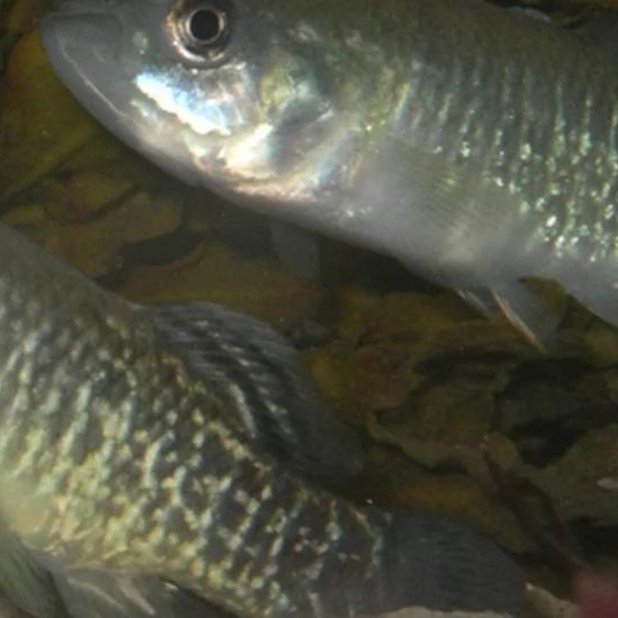
As fish migrate from fresh-water rivers to the salt-water ocean they must adapt to their new surroundings by excreting the excess salt that enters their bodies. This is done by a special type of cell that is present in the gills and operculum.
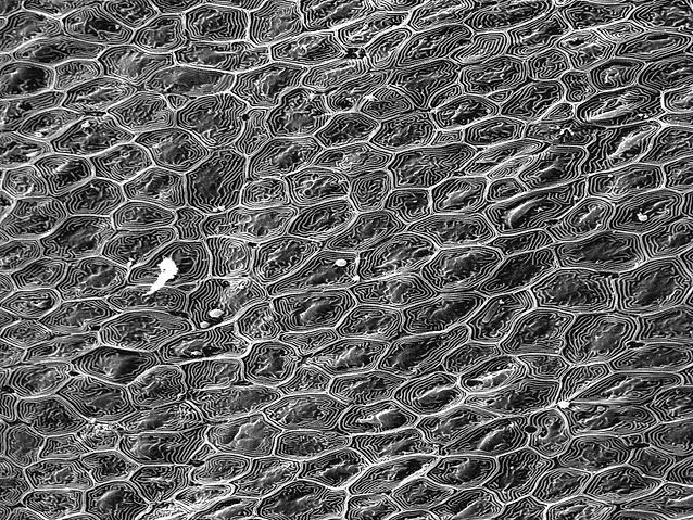
The image to the right is a scanning electron micrograph of the top portion of the cells covering the operculum.
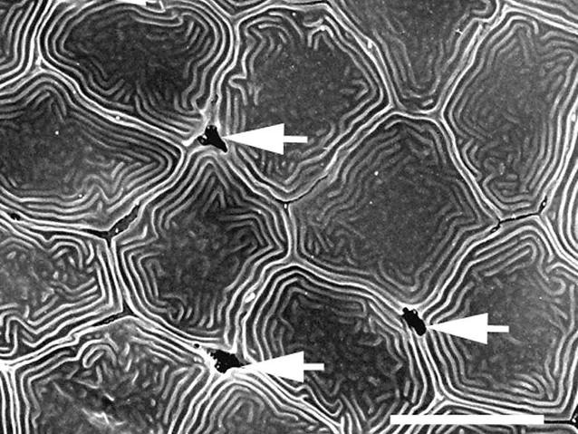
If we look closer, we can see that there are small gaps between the junctions of the opercular cells. These are indicated by the white arrows on the image to the left. It is through these gaps that excess salt is secreted back into the ocean.
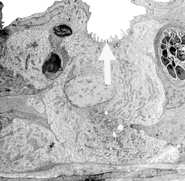
The special cell that performs this excretion is called an ionocyte. It is shown here in a transmission electron micrograph to the right. It uses a large amount of energy to excrete the salt through the gap (shown by the white arrow) and back into the ocean.
Contact
University Microscope Facility
@email
902-867-3841
J. Bruce Brown Hall
2320 Notre Dame Avenue
Antigonish, NS
B2G 2W5
