Explore the Microscopes
Learn more about the microscopes availble at the StFX University Microscope Facility.
TEM
Philips 410 Transmission Electron Microscope
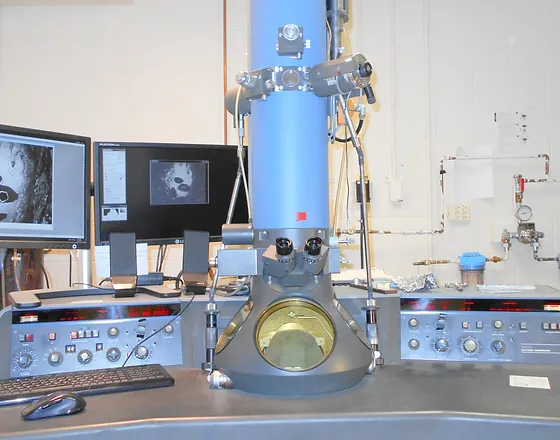
The TEM transmits a beam of electrons through a small piece of animal or plant tissue allowing the biologist to conduct a detailed investigation of the cells and the interior cellular components present. This powerful instrument can magnify details over 100,000 times and is an excellent tool for experimental scientists to see some of the smallest features of animal and plant cells.
*add links*
Click here to discover more projects the TEM has been used in.
Click here to view a gallery of TEM images taken at the UMF.
SEM
Jeol JSM 5300 Scanning Electron Microscope
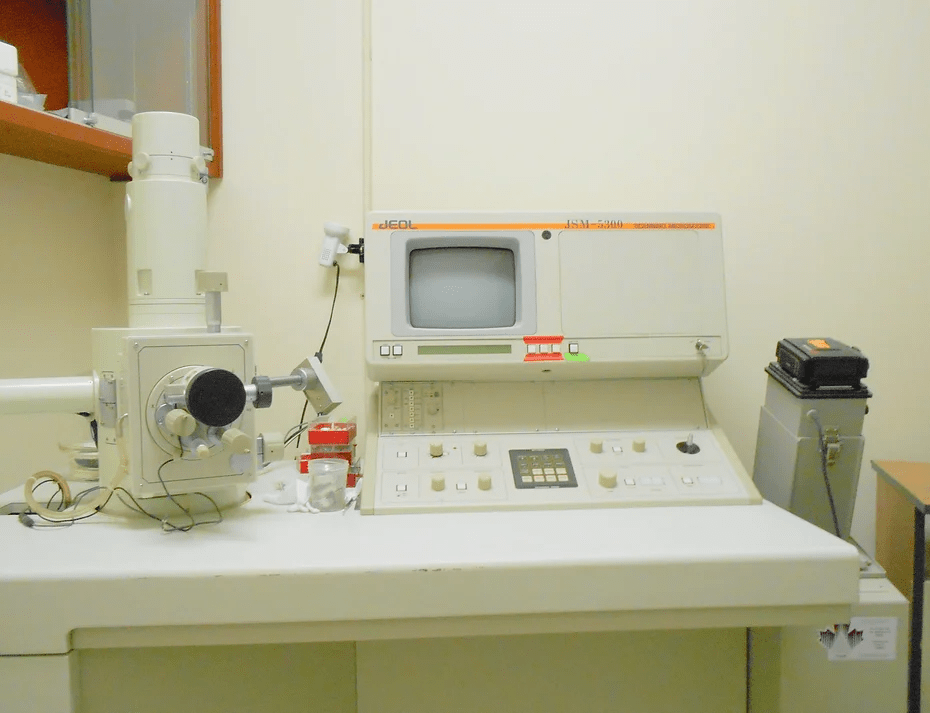
The scanning electron microscope scans a beam of electrons across the external features of small animals or tissue, collects this information, and then magnifies it up to 50,000 times. It is used to identify the surface features of insects, pollen and eggs. As the electron beam interacts with metal, the specimens must first be coated with gold!
*add links*
Click here to discover more projects the SEM has been used in.
Click here to view a gallery of SEM images taken at the UMF.
Confocal
Olympus Confocal Microscope
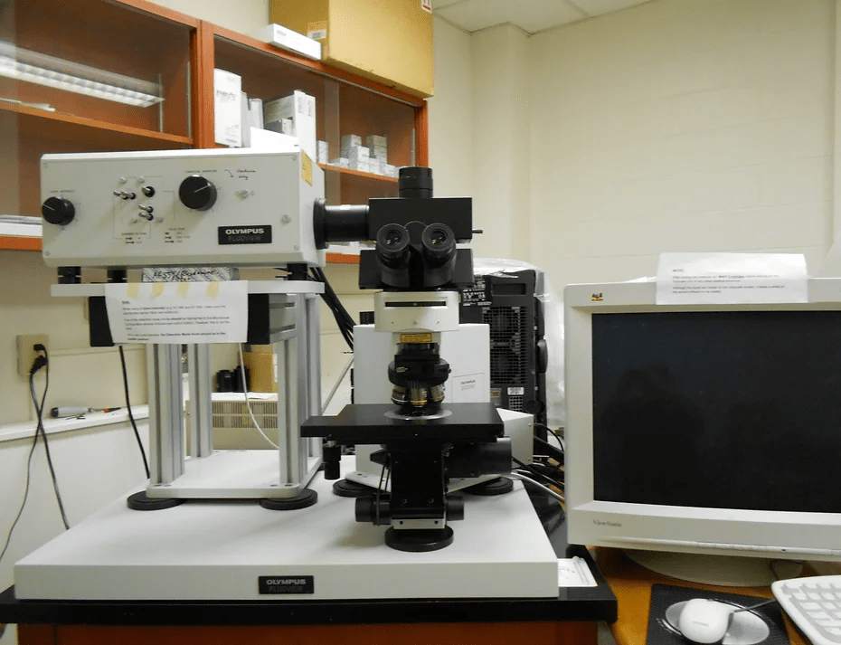
The confocal microscope uses lasers to excite fluorescent stains within biological tissue and can tell researchers about the molecular structure of their tissue of interest.
*add links*
Click here to discover more projects the Confocal has been used in.
Click here to view a gallery of Confocal images taken at the UMF.
Contact
University Microscope Facility
@email
902-867-3841
J. Bruce Brown Hall
2320 Notre Dame Avenue
Antigonish, NS
B2G 2W5
